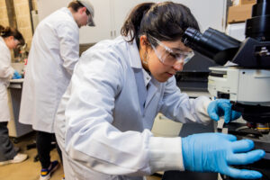
The biomedical engineering program received a boost this semester with the launch of the biomaterials and tissue engineering laboratory.
It’s the first “wet lab” at NIU’s College of Engineering and Engineering Technology (CEET), referring to experiments that are conducted on liquids, chemicals and biological matter.
The cutting-edge lab is equipped for growing and studying mammalian cells, the complex cells that make up the tissues and organs of mammals. It can also extract and use animal tissue materials to create scaffolds, specially designed material that helps cells grow and build new, healthy tissue for medical treatments.
Research in the laboratory builds on the college’s emphasis of bridging theory with practice, said Dave Grewell, CEET dean.
“This brings us to a whole new level of connecting our students with industry, national labs, and healthcare,” Grewell said. “The lab grows our ability to provide students real-world experience in biomaterials, tissue engineering, and medical devices—great preparation for careers in academia, industry or regulatory science.”
The lab includes several pieces of cell culture and analysis equipment:
- A biosafety cabinet, which provides a sterile environment for safe mammalian cell culture handling.
- A cell culture incubator, which maintains optimal temperature, humidity and carbon dioxide levels for cell growth.
- Multiple types of microscopes for analyzing cell morphology and scaffold-cell interactions.
- A microplate reader, which allows for measuring chemical changes and testing cell safety.
In addition, the lab has biomaterials and fabrication equipment:
- An oven for controlled heating of biomaterials for scaffold preparation.
- A ductless fume hood, which provides a safe environment for handling chemical processes involved in biomaterial fabrication.
- A freeze dryer, which helps dry and stabilize biomaterial scaffolds.
In January, the lab began operations under the guidance of Assistant Professor Hyung Jin Jung.

“These technologies allow students to gain hands-on experience in biomedical research by engaging in cell culture, biomaterial processing, scaffold fabrication and quantitative analysis,” Jung said. “This lab will be a huge asset to build students’ technical and problem-solving skills.”
Two teams of undergraduate students have begun using the lab for their senior design projects. Both teams are working on collagen scaffold fabrication and organ-on-a-chip development, engaging in both conceptual design and hands-on prototyping.
Jacob Hanson, who already has a job lined up as biomedical technician after graduation in May, said the lab “is much better than what we’ve had to work with before.”
“We have more resources and this is a more organized layout,” Hanson said. “And there’s a lot of potential here that we haven’t touched yet. It’s going to be even better for students in the future.”
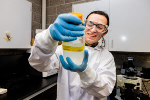
Among his three senior design teammates is Khoa Nguyen, who is working with Hanson on a 3D printing method to mold collagen, then using a freeze dryer in the lab to freeze it.
He echoed Hanson’s perspective: “It’s been a very good experience overall,” said Nguyen, who is also slated to work in the biomedical field after graduation. “We’re able to work with multiple pieces of equipment as we learn about safety, including how to dispose of hazardous materials.”
Student research in the lab will focus on:
- Mechanobiology– Studying how physical forces affect cells and using that knowledge to help grow and repair tissues.
- Biomaterials Development– Designing and fabricating scaffolds and medical devices for tissue reconstruction.
- Organ-on-a-Chip– Creating tiny lab-on-a-chip devices that imitate the human body to study diseases and test treatments.
Students and faculty will conduct experiments related to cell culture, biomaterial processing, scaffold fabrication, and characterization.
The organ-on-a-chip technology replicates physiological systems for disease modeling, drug testing, and studying intercellular interactions.
“This system helps analyze tissue responses to treatment, improving preclinical testing,” Jung said. “By offering a controlled environment to evaluate pericellular conditions and optimize treatment strategies, this technology could reduce reliance on animal studies and streamline human clinical trials, saving time and money.”
The lab is an integral part of BME336: Biomaterials, a course in which students conduct biomaterial processing, biodegradation assessments and biocompatibility evaluations.
The lab will also foster more interdisciplinary collaboration within and even beyond CEET, such as mechanical engineering and biology, to engage in biomedical research, Jung said.
One goal is to expand research to develop surgical techniques and implantable medical devices for preclinical animal studies, added Jung, who is recruiting BS/MS research assistants.
Learn more about the biomedical engineering program.
